Diagnostic Imaging
Many knee and hip conditions, and their resulting treatment, will require diagnostic imaging to identify the root cause of the problem. A diagnostic scan is a type of test ordered by medical professionals to help them to diagnose patients conditions, check how effective certain treatments are and plan appropriate treatment.
All diagnostic tests will be carried out at the same location as any potential surgery. You will be provided with all the information you need prior to your visit, including any special instructions required to prepare for your particular scan, what to bring on the day and how to find us.
One Stop Clinic
Mr Atwal currently runs a One Stop Clinic each Friday morning at the Nuffield in Cheltenham where in one visit patients will have:
• An initial consultation with Mr Atwal
• Any diagnostic imaging that you may require (MRI, X-ray, CT
scan)
• A report on the imaging by a Consultant Radiologist
• A follow up consultation with Mr Atwal to discuss the results
The entire process is all completed within approximately 2 hours, saving you multiple visits to the hospital. Following this, Mr Atwal will then work out a treatment plan with you to get the best results for your specific condition.
Common Diagnostic Tests
What is an MRI?
Magnetic Resonance Imaging (MRI) produces
high resolution pictures of any part of the
body in any direction. The scanner uses a
high-strength magnet, radio waves and
computers to generate images. MRI does not
use X-rays so it can be used repeatedly. It
is considered to be a very safe way of
producing images that can diagnose medical
conditions.
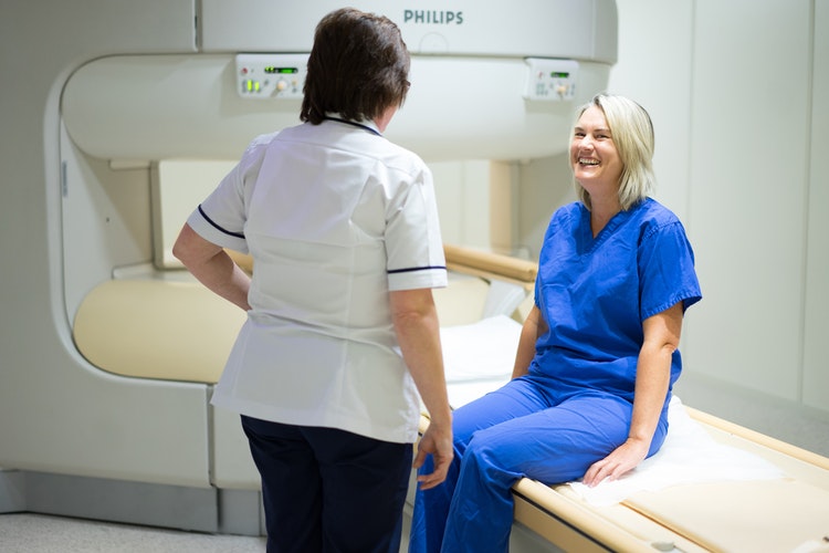
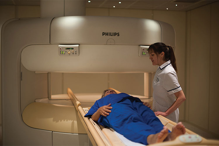
What to expect
The Radiographer will position you on a
comfortable couch, which slides into the
scanner. You will be asked to keep still
while we scan you and produce your images.
The scanner is very noisy when it is taking
pictures so we will provide you with ear
protection. Examples of the types of noises
you may experience during your scan can be
heard here.
There are no after-effects from your scan so you can carry on with all your normal activities straight away.
If you have a Buscopan injection, an injection into the joint during your procedure or have taken medications which make you feel relaxed or drowsy it is advised you are accompanied by someone who can drive you home.
How long will it take?
This depends on which part of the body is
being scanned. Most scans of one area are
completed in 20-30 minutes.
Results
Mr Atwal will receive your scan images and
report within 5 working days. You should
make an appointment with them to discuss the
results. The radiographer will not be able
to give you your results on the day of your
scan.
What is a CT scan?
The scan is carried out by a special type of
x-ray machine. The images the machine
produces are cross-sections of your body
(think slices in a loaf of bread). The parts
of your body can be shown in much greater
detail than in standard x-ray films, and
this helps the doctors diagnose your
condition much more accurately.
You will lie on a moveable bed and pass through the a ‘doughnut’ shaped scanner. A narrow, fan-shaped beam of x-rays is produced from inside the machine, which rotates 360 degrees around you. The x-rays pass through your body and are detected by electronic sensors on the other side of the machine. The information from the machine then passes to a computer which produces a picture of the structure of the inside of your body. The bed moves a small distance to position you for the next picture. It takes about a second to produce each slice. These pictures can then be reconstructed by the computer to form a complete image of the inside of your body.
Risks
CT scanning does involve being exposed to
radiation in the form of x-rays, but the
benefit of an accurate diagnosis far
outweighs the risk from the x-rays. However,
if you are pregnant, or may possibly be
pregnant, you must tell a member of staff
before the scan as unborn children are at
greater risk because they are still
developing. The amount of radiation used for
a CT scan is more than the amount used for a
standard x-ray and (depending on the
examination you have) is equal to the amount
of natural radiation you would receive from
the atmosphere over a period of around three
years. The CT scan itself is completely
painless.
Contrast injections and oral
contrasts
A contrast is a dye used to make blood
vessels and organs stand out in images.
Before the scan, you may have to take oral
contrast, have an injection of a contrast,
or you may have to have both. If you are
breastfeeding you should wait 24 hours after
a contrast injection before you breastfeed
again.
Can I bring a relative or
friend?
If you would like a friend or relative to
come with you, they may wait in the waiting
room.
At your appointment
When you arrive, please report to reception.
Once you have been signed in the
radiographer will come and explain the
procedure you are going to have. You should
tell them if you have diabetes, asthma, or
any allergies. If you need an oral (by
mouth) contrast the radiographer will give
you this before you go to the CT department.
If you need to remove any clothing someone
will show you to the changing rooms and give
you a patient gown or other special clothing
to change into.
During the scan you will be made comfortable on the moveable bed. Straps and pillows may be used to help you stay in the correct position and to help you stay still during the scan.
If you need an injection of contrast, you will be given this through a vein in your arm while you are lying on the scanner bed. This may make you feel warm and give you a metallic taste. The radiographer will stay with you during the injection. The radiographer will control the bed from the control room and will slowly move it to position the part of your body being investigated inside the ‘doughnut.’
The radiographers will be in the control room during the scan, but you will be able to talk to them using an intercom, and they will be watching you all the time. You will hear a clicking and whirring sound from the CT scanner during the procedure. During the scan the radiographer may ask you to hold your breath or to not swallow while each image is being produced – if you feel any discomfort or anxiety because of this, please tell the radiographer immediately.
What are x-rays?
X-Rays are a type of invisible
electromagnetic radiation – you don’t feel
them when they pass through your body. X-Ray
is used in many different ways to diagnose
medical conditions. An x-ray image is
produced when a small amount of radiation
passes through the body and hits a sensitive
screen placed on the other side. X-rays are
absorbed into tissues and bones in differing
amounts. For example, x-raying bone, which
does not allow much radiation to pass
through it, produces white images, whereas
x-raying lungs, which are less dense because
they are filled with air, produces darker
images.
Risks – what are the side
effects?
We are exposed to radiation from the
environment every day. Radiation is involved
in producing an xray but the dose you will
receive is very small – during an x-ray, you
will be exposed to around a fifth of the
radiation you would receive from the
environment over a year. This is similar to
the amount of radiation you would be exposed
to during a transatlantic flight. You should
tell your doctor or radiographer if there is
any possibility that you are pregnant –
x-rays are a greater risk to unborn children
because they are still developing.
Before your
appointment
You do not need to make any special
preparations for your X-Ray. However, please
remove any jewellery that may interfere with
the x-ray if you can, for example it would
be helpful if you could remove rings for
hand x-rays and necklaces for chest x-rays.
The x-ray procedure
The radiographer may ask you to sit, stand
or lie on the table depending on which body
part is being examined. The radiographer may
move you to get the correct position. The
radiographer will stand behind a screen
while they take the x-ray – although each
procedure only produces a small does of
radiation, these doses would begin to add up
for the radiographer if they had no
protection. Once the procedure is complete,
you will be asked to wait while the
radiographer checks the images. The actual
procedure usually only lasts five to 10
minutes.
Can you bring a relative or
friend?
Yes, but for safety reasons they may not be
able to go in the x-ray room with you.
The results
The radiographer will not be able to give
you any results on the day. A consultant
radiologist will report on the x-ray images
and send the report and images to Mr Atwal
who will then explain the results to you. In
many cases, you may take a CD of the x-ray
home with you on the day.
For more information on any diagnostic imaging please do not hesitate to get in touch with Mr Atwal through our appointments page.
QUICK ENQUIRY
